Getting started, tracing, analysis and specification of the obtained data
Medicom MTD. Medical equipment for electrodiagnosis, rehabilitation and research
Phones: +7 (8634) 62-62-42, 62-62-43, 62-62-44, 62-62-45.
www.medicom-mtd.com

Software for three-dimensional localization of electrical activity sources "Encephalan-3D"
An important EEG problem, particularly in epileptiform activity analysis, is localizing its focus (the position in three-dimensional coordinate system) as accurately as possible. Solution of this problem is based upon the idea that elevated pathological activity appears in the local brain lesions area influencing electric activity generation of the entire cerebral cortex.
The calculation of the coordinates of the possible location of the source of electrical brain activity (three-dimensional localization) is based on a mathematical model, in which the activity source is presented in the form of an electric dipole located at the locus center. Head size, inhomogeneous conduction of head tissues, non-spherical form of skull are all taken into account in calculation.
Software "Encephalan-3D" is designed for three-dimensional localization of the sources of abnormal electrical brain activity based on the analysis of scalp EEG/EP electrodes and is used as an auxiliary method, especially in cases where the focus of pathological activity has no obvious morphological changes and is not detected by computed tomography and MRI.
The advantage of this method is the high time resolution, which allows evaluating the EEG in the record (including long-term recordings) or EP dynamics of electric brain activity focus in different pathologies (often of epileptic characteristics).
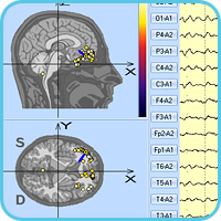
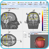
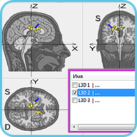
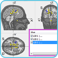
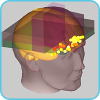
The equivalent dipoles calculated are shown as dots in diagrams of the three orthogonal projections of head in the Cartesian coordinate system (stereotactic one) with attachment to anatomic landmarks so that allows correlating them with the relevant tomograms of brain and attaching them to certain brain structures
Three-dimensional localization of sources of pathological electric brain activity by the "Encephalan-3D" application allows identifying the source of pathological EEG, defining its intensity and depth of occurrence, examining spatial (prevalence, any phase shifts etc.) and temporal features of its focus (stability of discharges, similarity of their parameters, and others).
Software "Encephalan-3D" is an optional software to:
Getting started, tracing
One or more EEG fragments with characteristic features are selected for staged localization of a source (tracing). The required EEG fragments can be selected in software for EEG studies ("Encephalan-EEGA" or "Encephalan-EEGR") or directly in "Encephalan-3D" Software. EP-studies data can also be downloaded for localization from "Encephalan-EP" software.
It is necessary to choose the localization model (single-, two- or three-dipole) to calculate the localization of the source. For this purpose the topography of potential distribution in several EEG time cuts is preliminarily evaluated, the number of the most typical and stable focuses is computed by EEG amplitude map.
During tracing, the software is alternatively analyzing EEG/EP time samples. Processing is displayed by moving of a special marker. Found dipoles are displayed on head cuts. At the same time, localization is analyzed for conformity so that a user can pause the tracing and adjust the settings in case of a large number of mismatches.
Analysis and specification of the obtained data
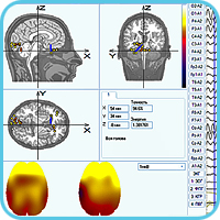
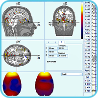
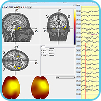
At the end of the tracing, the Localization window displays dipole "clouds" (groups) on the head cuts, two amplitude topographic maps based on the native EEG and localization data (for quick visual comparison and evaluation of the result), and a table with coordinates, accuracy and energy values of the selected dipoles.
Any head projection with found dipoles can be viewed enlarged, as well as dipoles distribution over three-dimensional head model, and graphs of temporal and spatial localization dynamics.
An important feature of "Encephalan-3D"┬╗ software is the simultaneous displaying of changes in all displayed data: dipoles distribution, topographic maps and localization graphs for a particular moment in time are displayed when moving the marker over the EEG signal. And vice versa, when specifying a particular dipole, the marker on signals moves to EEG cut, where the dipole has the maximum energy, and the table displays its characteristics, etc.
Localization of source is carried out both for one fragment only and several selected ones.
Specification of source for localization by several EEG fragments confirms the correct selection of EEG fragments and the location of the source of abnormal electrical activity
The "Encephalan-3D" application allows numerically estimating the accuracy of scalp voltages restoration by comparing the instantaneous amplitude values of initial and reconstructed signals of the current time cut, as well as visually comparing amplitude topographic maps.
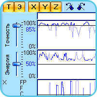
Calculation and display of accuracy and energy changing graphs, and spatial and temporal dipole motion path for all localized fragments are performed to estimate the source dynamics.
These graphs allow identifying dipole switch moments, for example in the presence of mirror foci.
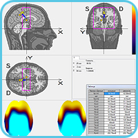
Numerical evaluation of the localization accuracy in the form of a table with initial and reconstructed data
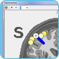
Since the great number of dipoles and their close location to each other on the screen, the Enlargement window is used for a detailed view.
Dipole of maximum accuracy and maximum energy (according to color scale) is of the most interest.
There can be displayed not only dipoles but also coordinating vectors with diagnostically significant dynamics on the enlarged head cut.
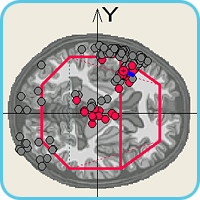
Setting and displaying specified spatial brain areas
For further analysis of dipoles found during localization, the application pro-vides setting of coordinates and sizes of the special three-dimensional areas, such as central, temporal or occipital. Data on dipoles, their parameters, and location in a particular spatial area are recorded for a detailed comparison or statistical analysis in ASCII or MS Excel format.
Localization correction taking into account the oculomotor artifacts
In certain cases, physiological signals of non-cerebral origin make a significant impact on the EEG and cause serious distortion of the three-dimensional localization results.
For example, attachment of two dipoles in the eyeball area allows compensating the effect of oculomotor artifacts on the localization calculation.
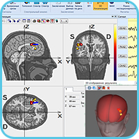
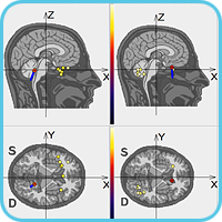
Three-dimensional localization of sources can be applied not only to typical EEG graph elements (e.g., spike-wave), but also to evoked potentials (EP) components. Cognitive EP P300 example shows localized regions that are located in the front areas of the brain and characterize the most active areas of the brain in endogenous processes of the relevant stimuli recognition.
Three-dimensional localization for EP data requires their export to UDF format (to "Encephalan-EP" Software) and further import to "Encephalan-3D" Software.
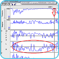
The first two images show the localization for averaged EP CNV responses (complex stimulus РђЊ a click, a pause, probabilistic stimulus: significant РђЊ 30%, insignificant РђЊ 70%) from 3 averagers: "Significant", "Insignificant" "General".
The last image shows localization analysis during the EP P300 study (changing the source location in reaction to significant and insignificant stimulus).
|
Phones: |
Frunze str., 68, Taganrog, |
| © 1997-2024 Medicom MTD Ltd All rights reserved | |

