ООО НПКФ «Медиком МТД»: Медицинское оборудование для диагностики, нейрофизиологии и реабилитации
Тел.: (8634) 62-62-42, 62-62-43, 62-62-44, 62-62-45.
www.medicom-mtd.com
The System of control and analysis of psychophysiological information – The System CAPI
(TU 26.51.66-039-24176382-2022, Declaration of Conformity of the EAEU N RU D-RU.PA04.B.53707/22)
Wireless amplifier units for models of the System CAPI
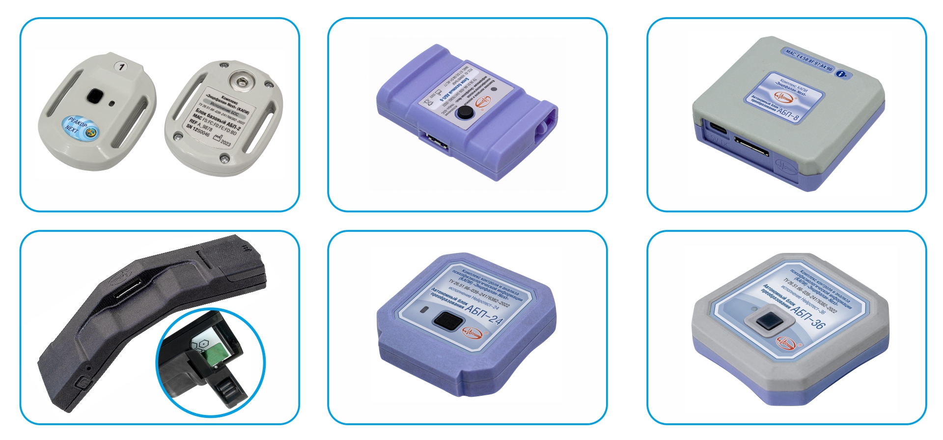
The wireless amplifier unit ABP-2 is the main unit for the Egoscop-Next and Rehacor-Next models of the System CAPI. The unit has one Micro-8M type input connector for two bipolar or two monopolar EEG derivations or two polygraphic sensors/electrodes and has an I2C interface input for digital sensors.
The wireless amplifier unit ABP-6 is the main unit for the model of the NFB System CAPI. The unit provides EEG recording of up to 8 (including derivation A1-N) monopolar derivations. ABP-6 has a built-in motion sensor and multifunction button.
The wireless amplifier unit ABP-8 is the main unit for the model of the BFB-Universal System CAPI and provides EEG recording from 8 monopolar or 8 bipolar derivations. ABP-8 has a built-in motion sensor and multifunction button.
The wireless amplifier unit ABP-24-AT is the main unit for the model of the Encephalan-Next-24-AT System CAPI. The unit provides EEG recording using 20 or 22 monopolar EEG derivations. ABP-24-AT has a built-in motion sensor and multifunction button, the ability to autonomously record a signal and a removable battery.
The wireless amplifier unit ABP-24 is the main unit for the model of the Encephalan-Next-24 System CAPI. The unit provides EEG recording from 20 or 22 monopolar EEG derivations, including from the reference channel A1–A2, as well as ECG from 1 channel and EOG from 2 additional channels. ABP-24 has a built-in motion sensor and multifunction button.
The wireless amplifier unit ABP-36 is the main unit for the model of the Encephalan-Next-36 System CAPI. The unit provides EEG recording from 32 monopolar EEG derivations, including from the reference channel A1–A2, as well as ECG from 1 channel and EOG from 2 additional channels.
Neuroheadsets for wireless compact wireless amplifier units (ABP)
for EEG recording from the System CAPI
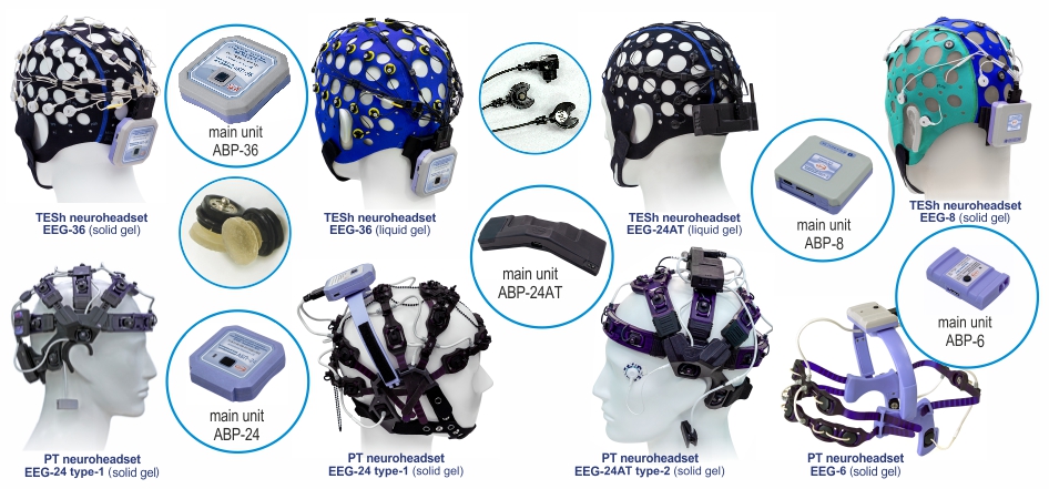
TESh neuroheadsets for ABP-36 main unit provide EEG recording using both snap electrodes for solid gel (left) and electrodes for liquid gel (right). The conductors are assembled into cables with a common ST40-36 type connector for connection to the ABP-36 main unit.
Silver chloride removable electrodes for liquid gel.
TESh neuroheadset (top) and PT neuroheadset (bottom) provide EEG recording both using electrodes for liquid gel and using snap electrodes for solid gel. The conductors are assembled into cables with a common ST40-24 type connector for connection to the ABP-24-AT main unit.
The TESh neuroheadset provides EEG recording using snap electrodes for solid gel. The conductors are assembled into cables with a common ST40-24 type connector for connection to the ABP-8 main unit.
Snap silver chloride electrodes with solid gel.
PT neuroheadsets (two types) provide EEG recording using snap electrodes for solid gel. The conductors are assembled into cables with a common ST40-24 type connector for connection to the ABP-24 main unit.
The PT neuroheadset provides EEG recording using snap electrodes for solid gel. The conductors are assembled into cables with a common ST40-10 type connector for connection to the ABP-6.
Connectors for ABP units for stationary use, EEG electrodes, electrode systems
and neuroheadsets from the System CAPI
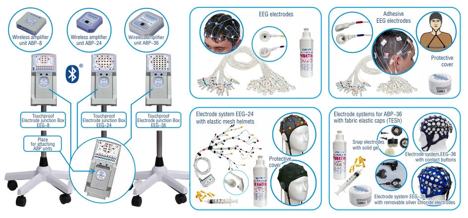
The connectors are used for EEG recording using standard electrodes with touchproof connectors in monopolar or bipolar derivations, providing electrical connection to the amplifier input jacks on the connector panel through the group connector of the main unit located inside the connector.
The connector lid allows you to install and remove the ABP main unit from the connector.
The connection of the amplifier inputs with the connector sockets for touchproof EEG electrodes is provided by a cable located inside the connector.
On the back side of the connector there are fastenings for VESA-100 type brackets, and there is also a compartment with a lid to accommodate the contactor of the USB power supply adapter from the sale package according to TU 9441-023-24176382-2008 for external power supply of the ABP main units from the USB interface socket located in PC, or in a network adapter with a USB connector.
Electrodes for contact gel with fixation by silicone flagellated helmets. Used for routine EEG studies.
Included: EEG cup electrodes, including 3 spare ones; set of EEG electrode clamps “ear clip”; a set of conductors (with a snap) for disposable ECG, EOG or EMG electrodes.
Adhesive EEG electrodes for use with connectors or cable adapters for long-term EEG monitoring.
They are distinguished by more reliable fixation of electrodes and high-quality EEG recording. Used for long-term EEG monitoring, EEG/PSG studies, neuromonitoring and for recording brain death.
Used with the wireless amplifier unit ABP-24. The electrode conductors are assembled into a common cable and have a group connector for connection to the ABP unit.
Recording of 20 EEG derivations is provided (14 derivations for ES-EEG-13-3G), 2 EOG derivations, 1 EMG derivation, 1 non-standard ECG derivation (one ECG electrode relative to the reference EEG electrode).
Electrode systems with fabric elastic caps (TESh) provide EEG recording of up to 36 monopolar derivations (including derivation A1-N).
The sensor enclosures of snap EEG electrodes are cup-shaped. At the base of the sensor enclosure there is a silver chloride current collector, which is connected to a snap on the outer surface of the cup. Solid gel inserts are attached in the electrode cup.
Additional devices from the System CAPI
Eye tracker glasses ATV-2-200.
TU 26.51.66-040-24176382-2023
Eye tracker glasses ATV-2-200 and TESh neuroheadset with EEG wireless amplifier unit ABP-36.
Eye tracker ATV-1, modification ATV-1K (TU 26.51.66-035-24176382-2020) provides control and analysis of oculomotor reactions when the client works with the presented visual content as part of the System synchronously
with recorded physiological signals. This ensures data transfer from the eye tracker to the System PC via a LAN in the form of LSL streams. Purposed for desktop use in conjunction with a 24-inch monitor.
Eye tracker ATV-1, modification ATV-1C (TU 26.51.66-035-24176382-2020), unlike the ATV-1K model, is manufactured for the floor-mounted variant of use in conjunction with TVs or a monitor with a large screen
(40, 55 or more inches in size diagonal), as well as with a large screen and video projector (with a size of 1, 2, 3 or more meters), while the eye tracker is placed on a special stand that provides the required angle of inclination of the eye tracker and its height relative to the client’s head, depending on the size of the screen, on which visual information is presented. The ATV-1C model also has the original ability to automatically determine the spatial position of the tracker relative to the large screen using a built-in rear view camera.
Additional modules and devices from the System CAPI
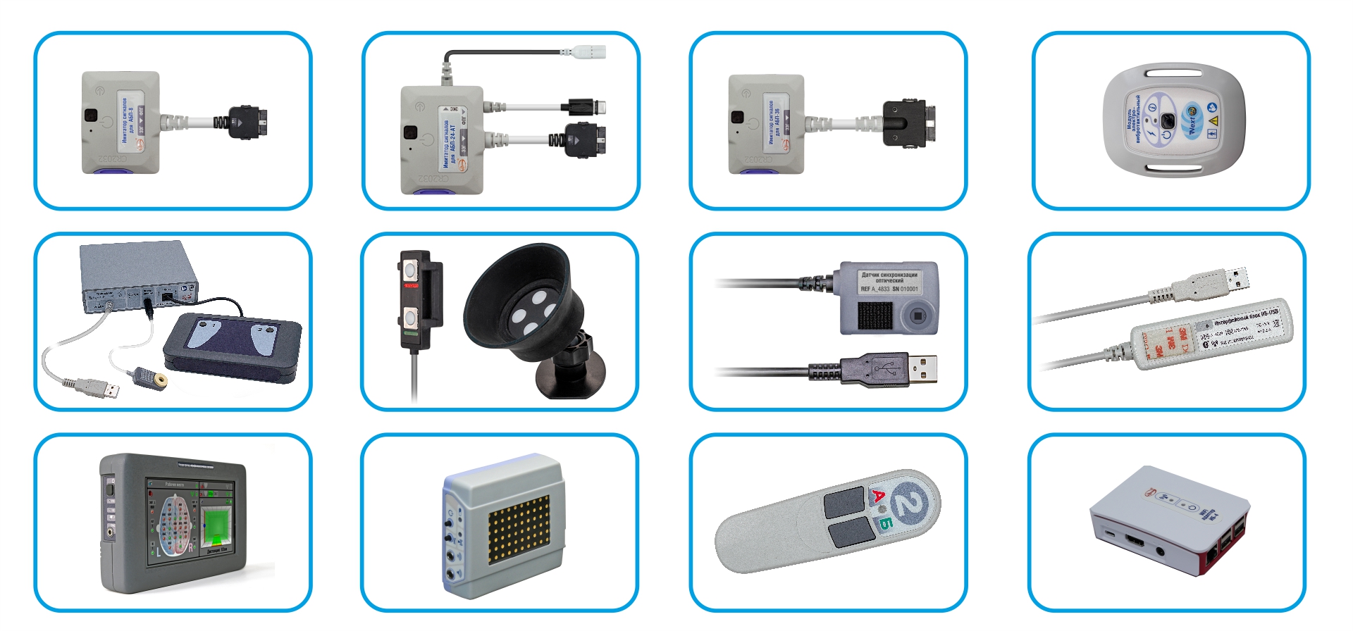
Simulator IS-8 has a cable with a 24-pin connector (ST40X-24S), which is connected to the corresponding socket of the ABP-8 or ABP-8-I unit.
Simulator IS-24-AT for the ABP-24-AT unit. The composition includes cables for checking EEG channels, for checking the ECG channel and with a type-C connector for checking the digital PPG channel.
Simulator IS-36 has a cable with a 36-pin connector (ST40X-36S), which is connected to the corresponding socket of the ABP-36 unit.
Module electro-vibrotactile (MEV) generates an electrical or vibrotactile information stimulus to the client being tested through built-in electrodes or a vibration source.
Primebox Device is used to measure reaction time and provides recording of the client's reaction time. The device is connected to the USB port of the System PC.
Attention Reserve Control Device provides the client with stimuli from red and green LEDs and records the instructor’s reaction at the press of a button.
Optical Synchronization Module captures the video synchronization stimulus on the PC monitor screen using a built-in optical sensor and a Bluetooth 5.0 wireless module.
The Wireless PC adapter IB-USB provides connection between the PC and the main ABP units, as well as modules and devices of the System via 10 information channels via the Bluetooth 5.0 interface.
Wireless concentrator of neurophysiological signals IB-KNS provides display of information about data from wireless modules and devices on the touch screen.
Wireless stimulator SFN/FO-04 is used to study auditory, visual and somatosensory long-latency evoked potentials in psychophysiology.
Button sensor with two buttons for events, a built-in LED and a vibration module for presenting stimuli, as well as a built-in movement activity sensor and a Bluetooth 5.0 module.
Wi-Fi Adapter (for IB-4 and IB-USB) is purposed to increase the distance of the System equipment from the PC through the use of an adapter with a Wi-Fi interface between the PC and IB-4 or IB-USB.
Wireless units and signal recording modules from the System CAPI
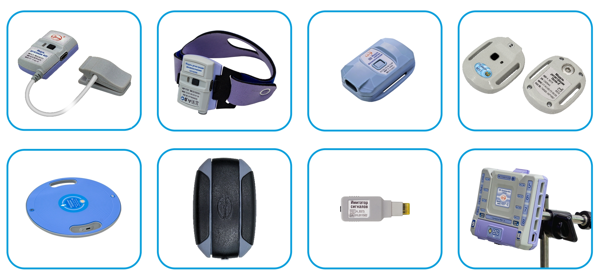
EarPPG module. The module has a built-in PPG channel (sensor on the ear lobe) and a “Bluetooth 5.0” wireless module.
Wristband for physiological signals monitoring (PS module). The module has built-in PPG, SkC, movement activity channels and an additional ECG channel, as well as a built-in “Bluetooth 5.0” wireless module and a marker button.
Electrophysiological module. The module has two sockets on the back for adhesive snap electrodes EEG, ECG, EMG, as well as a type-C connector for the I2C digital interface, a built-in movement activity sensor and a “Bluetooth 5.0” wireless module.
Wireless amplifier unit Poly-2. The module has one Micro-8M type input connector for two printing channels, providing connection of sensors and electrodes from the System. The I2C digital interface pins go to the same connector.
Wobble platform. Wobble platform has a built-in movement activity sensor and a “Bluetooth 5.0” wireless module.
Muscle tension module (tensometric). The module provides registration of muscle activity of the hands using a built-in movement activity sensor and the “Bluetooth 5.0” module.
Simulator IS-Micro. Simulator has an 8-pin EEG connector (Micro-8M) and is connected to the corresponding socket of the ABP-2 unit or wireless amplifier unit Poly-2.
Junction Box Poly-8. The operation of the main unit ABP-8 located inside is ensured. Provides 8 inputs for micro-8 polygraph sensors and electrodes, and 2 inputs for digital sensors and I2C devices.
Combined sensors from the System CAPI for recording physiological signals
via polygraphic channels
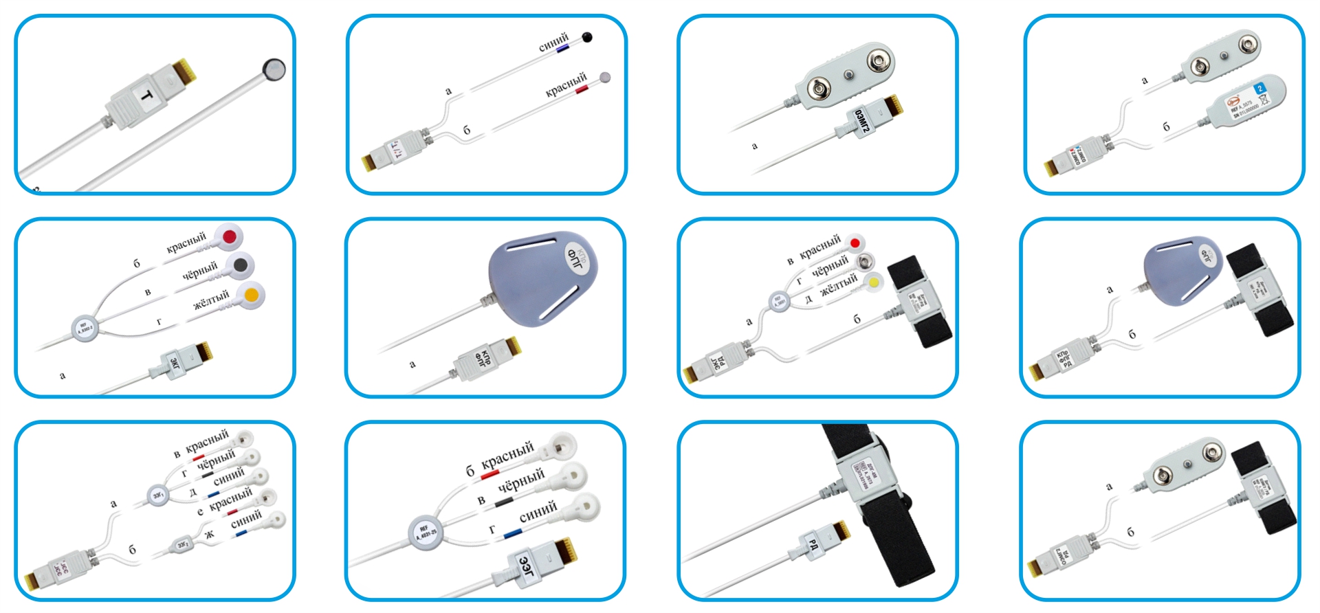
Temperature T* Sensor (Micro-8M). To assess the skin surface temperature of a selected part of the body. Has a low measurement time constant.
Combined T*/T* Sensor (Micro-8M). To record temperature from two sensors (for example, forehead - solar plexus, left hand - right hand).
Envelope EMG Sensor (double) (Micro-8). To assess the tone of a selected muscle based on envelope EMG measurements. The sensor enclosure has a contact with the N electrode function.
Combined EEMG2/EEMG2 Sensor. To record the tone of symmetrical muscles, assessing asymmetry, synergy and reciprocity.
ECG-Cable (with N electrode) (Micro-8). For recording bipolar ECG derivations with electrodes of various types.
Combined SkC/PPG* Sensor. (Micro-8M). For recording the parameters of the tonic and phasic components of the SkC and PPG parameters.
Combined ECG/RespEf Sensor. (Micro-8M). For recording of ECG and RespEf in order to assess the degree of cardiorespiratory resonance, the balance of the part of ANS.
Combined SkC/PPG*+RespEf Sensor. (Micro-8M). For recording the parameters of the tonic and phasic components of the SkC and PPG parameters.
Combined EEG/EEG Sensor, (Micro-8M). EEG recording in 2 symmetrical bipolar derivations using adhesive cup electrodes.
EEG Bipolar Cable (Micro-8). For EEG recording.
Respiratory Effort Sensor (Micro-8). To assess the parameters of abdominal or thoracic breathing (frequency and amplitude of breathing, duration of the inhalation and exhalation phases).
Combined EEMG2/RespEf Sensor (Micro-8M). For recording breathing parameters (the ratio of thoracic and abdominal) and assess muscle tension.
Combined sensors from the System CAPI for recording physiological signals
via polygraphic channels
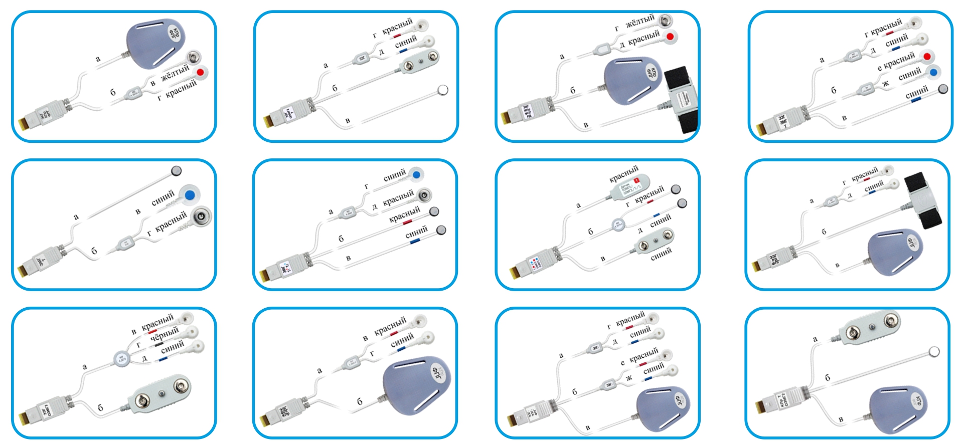
Combined ECG/SkC/PPG* Sensor (Micro-8M). For recording ECS, SkC and PPG in order to assess the degree of activation of the ANS based on HRV, SkC and vascular tone.
Combined EEG/EEMG2/T* (Micro-8M).
Combined ECG/SkC/PPG*+RespEf* Sensor, (Micro-8M). For recording physiological parameters to assess the actual PPS.
Combined EEG/EMG/T* Sensor (Micro-8M). For recording the temperature of the limbs and muscle tone of the facial muscles.
Combined EMG/T* Sensor (Micro-8M). For control the temperature of the limbs (finger) and muscle tone of the frontal muscles.
Combined EMG/T*/T* Sensor (Micro-8M). For recording the temperature of the fingers and muscle tone of the frontal muscles.
Combined EEMG2/EEMG2+T* +T* Sensor (Micro-8M). For recording the tone of symmetrical muscles, assess asymmetry, synergy and reciprocity.
Combined SkC/PPG*+EEG+RespEf* Sensor (Micro-8M). For recording physiological parameters that provide multimodal BFB training.
Combined EEG/EEMG2 Sensor (Micro-8M). For recording physiological parameters that provide multimodal BFB training.
Combined SkC/PPG*+EEG Sensor (Micro-8M). For recording physiological parameters that provide multimodal BFB training.
Combined EEG/EEG+PPG* Sensor (Micro-8M). For recording physiological parameters that provide multimodal BFB training.
Combined SkC/PPG*+EEMG2+T* Sensor (Micro-8M). For recording temperature, SkC and EEMG during stress resistance BFB training.
Combined sensors from the System CAPI for recording physiological signals
via polygraphic channels
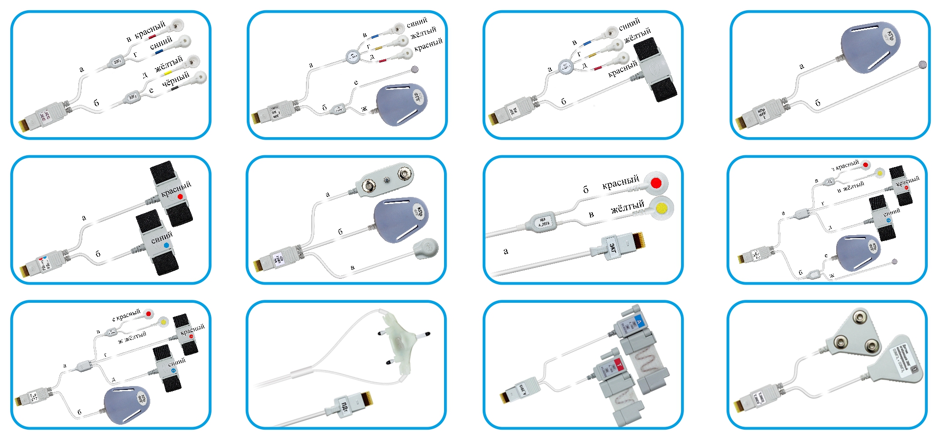
Combined EEG/EEG Sensor (Micro-8M). For recording EEG in derivations in accordance with the scheme.
Combined EEG/EEG+PPG*+ T* Sensor (Micro-8M). For recording parameters in accordance with the scheme for neurofeedback.
Combined EEG/EEG+ RespEf* Sensor (Micro-8M). For neurofeedback.
Combined SkC/PPG*+T* Sensor (Micro-8M).
For biofeedback.
Combined RespEf/RespEf Sensor (Micro-8M). For recording breathing parameters, the ratio of the amplitude of thoracic and abdominal breathing.
Combined SkC/EEMG2/Move* Sensor (Micro-8M). For recording tremor and movement activity.
ECG-Cable (Micro-8). For ECG recording. An N electrode is required.
Combined ECG/SkC/PPG* +RespEf*+RespEf*+T* Sensor for assessing the degree of activation of the ANS by HRV, SkC and vascular tone, as well as to record breathing parameters and temperature.
Combined ECG/SkC/PPG* +RespEf*+RespEf* Sensor for assessing the degree of activation of the ANS by HRV, SkC and vascular tone, as well as to record breathing parameters.
Oro-Nasal Airflow Sensor (Thermistor) “T-Flow” purposed to assess breathing flow parameters. Can be used in conjunction with a nasal breathing flow cannula.
Combined Abdominal and Thoracic RIP Sensors. For recording breathing parameters, the ratio of the amplitude of thoracic and abdominal breathing.
Combined EEMG3/EEMG3 Sensor is purposed to record the tone of symmetrical muscles, assessing asymmetry, synergy and reciprocity.
|
Phones: |
Frunze str., 68, Taganrog, |
| © 1997-2024 Medicom MTD Ltd All rights reserved | |
